
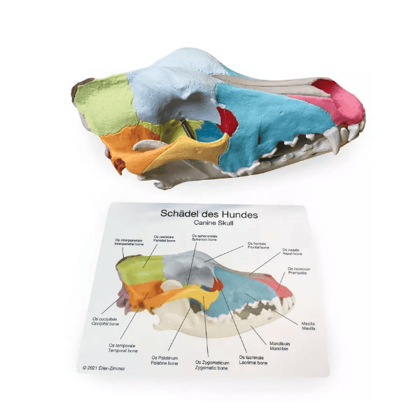
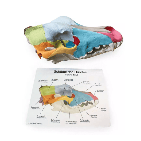
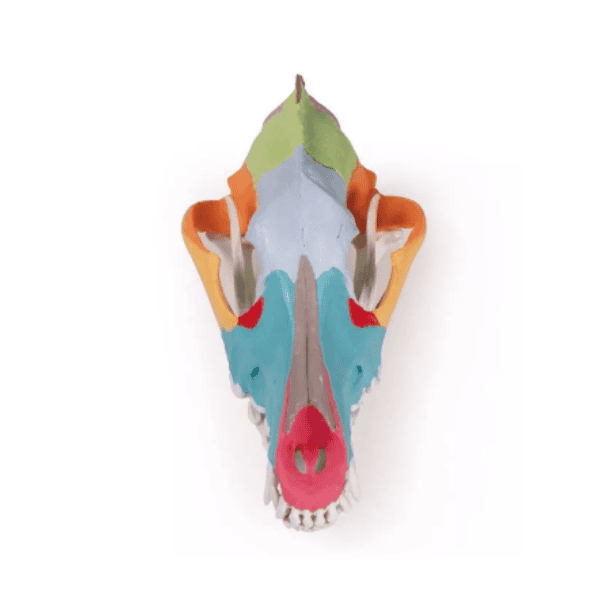
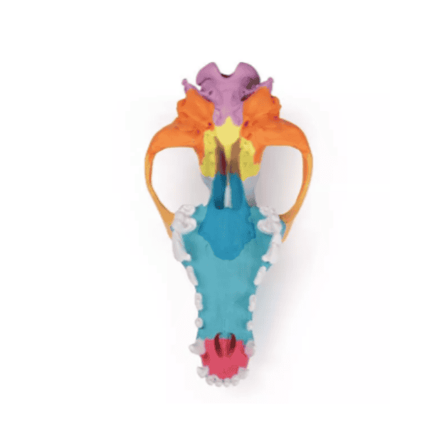
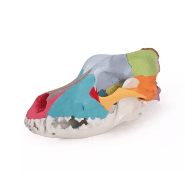
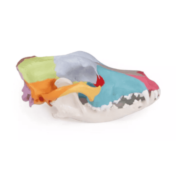
Dog Skull with Didactic Painting
SKU:VT028074
Related Products
Make Complex Skull Anatomy Easier to Identify
The number of closely connected bones within the canine skull can make identification challenging when students first encounter cranial anatomy. Didactic coloring visually differentiates individual bones, helping learners recognize boundaries and understand how structures relate within the complete skull.
Students can rotate and examine the model from multiple perspectives while comparing structures with anatomy texts, diagrams, or imaging. This three-dimensional view is particularly useful for studying areas that can be difficult to interpret from a flat illustration alone.
As students move beyond basic identification, the model provides a useful anatomical foundation for dentistry, diagnostic imaging, surgery, and examination of the canine head.
Product Images
Stay Up to Date on Products
Get Emails from VetiSimLab